RFA For Live logo

Thyroid Health

Thyroid Ultrasound Trilogy – I. The Basics of Ultrasound

Have questions about your thyroid ultrasound? We are here to answer!

Inspired by some patients’ requests, we have prepared a series of Thyroid Ultrasound articles to help you better understand those terminologies on your report and ask the right questions during your appointment. In this first article, let’s start with some basics of ultrasound.

Diagnostic ultrasound is a non-invasive imaging modality that uses non-ionizing high-frequency sound waves to produce images of your internal structures. The ultrasound transducer emits a sound beam that propagates through the tissue then waits for the returning echo reflected from the structures in the plane (path) of the sound beam, creating a grey-scale image we see on the screen.
Diagnostic Ultrasound

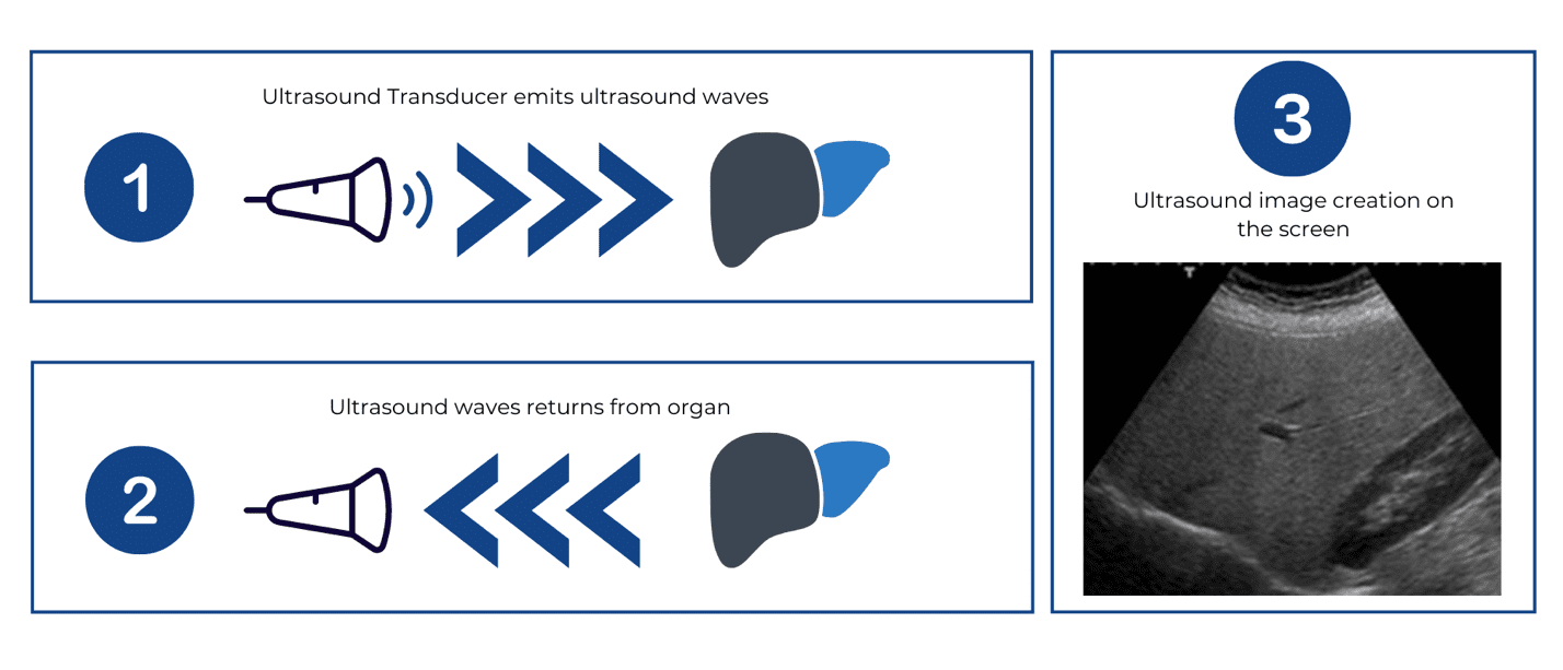
Brightness Mode (B-Mode)

ultrasound is most frequently used in thyroid ultrasounds supplemented by Color Flow Doppler to visualize vascularity on the gland. Depending on the strengths of the returning echo, the echo is registered as dots of various brightness levels to produce the greyscale image that we see on the screen. A structure that returns a stronger (higher-amplitude) echo will display a brighter dot than a structure that returns a weaker (low-amplitude) echo.

However, the appearance of the structure’s brightness on the screen depends on its surrounding environment. In order for a structure to appear salient on ultrasound, contrast is pertinent. It is similar to the differences that you observe when you draw on a blank paper using a white crayon versus black crayon.

Structures that generate stronger echo relative to its neighboring tissues will appear brighter on the screen and vice versa. Depending on the sound attenuation (loss of sound along its path), the amplitude (strength) of returning echo of different tissues varies. Physicians and sonographers can use the varying brightness levels of tissues to characterize tissue composition.
Liver Parenchyma
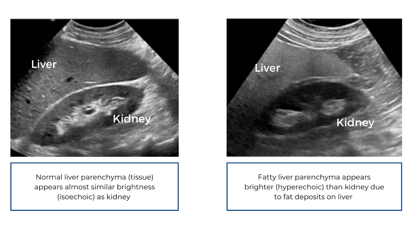
Now, we’ve learned the basics of ultrasound physics, in the next article we will go over some common terminologies used in ultrasound.

Sanders, R. C. (2016). Clinical sonography: A practical guide. Philadelphia: Wolters Kluwer.

Rumack, C. M., Wilson, S. R., & Charboneau, J. W. (2005). Diagnostic ultrasound. St. Louis: Elsevier Mosby.

2 Responses

Leave a Comment

Your email address will not be published. Required fields are marked *

More From Our Blogs

Why Might Thyroid Nodules Need To Be Surgically Removed?

Thyroid nodules can be diagnosed as two primary forms, malignant (cancerous) or benign (non-cancerous). This diagnosis is one of the most significant factors considered before a suitable method of treatment is prescribed. Is it necessary

Important Safety Information / Disclaimer
Patients should talk to their doctor to decide if STARmed Thyroid RFA is right for them. Patients and doctors should review all available information on non-surgical and surgical options and associated risks in order to make an informed decision. Individual results may vary, testimonials are not claimed to represent typical results. All testimonials are from real patients, and may not reflect the typical patient’s experience, and are not intended to represent or guarantee that anyone will achieve the same or similar results.
The information on this site is not intended or implied to be a substitute for professional medical advice, diagnosis or treatment. All content, including text, graphics, images and information, contained on or available through this web site is for general information purposes only.
Registered trademarks of TaeWoong Medical USA or their respective owners.

Indication
The VIVA Combo RF Ablation System and star RF Electrode are intended for use in percutaneous and intraoperative coagulation and ablation of tissue.

Contraindications
There is a risk that error may result due to the radiofrequency current on patients who have pacemakers and other active implants. Do not use the radiofrequency lesion generator and electrode on these patients.
Complications:
The following types of complications may result due to the use of the radiofrequency lesion generator and electrode.
– Tumor recurrence
– Burn due to the over-heating of the the surgical equipment
– Dangerous situation due to the unskilled equipment control
– Cross-infection or complications due to the re-use of the inappropriate electrode
– Ascites/diarrhea
– Bleeding of the coagulated part
– Ventricular fibrillation
– Weakness of liver functions
– Symptoms after RFA treatment includes (abdominal) pain, fever, nausea, headache, right shoulder joint pain and chest discomfort might occur

RFA for Life is committed to protecting your privacy under the relevant laws in the USA, and we’ll only use your personal information to administer your account and to provide the products and services you requested from us.
By clicking submit below, you consent to allow RFA for Life to store and process the personal information submitted above to provide you with the contents requested.

Physician Finder

You are taking a big step by exploring your treatment options. You are not alone. Many other people were searching for the same answers too. Many of these individuals chose RFA.