RFA For Live logo
Thyroid Health

Thyroid Ultrasound Trilogy – III: How Does Your Thyroid Look Like on Ultrasound

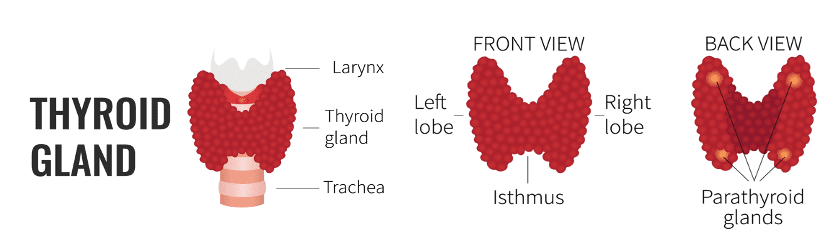
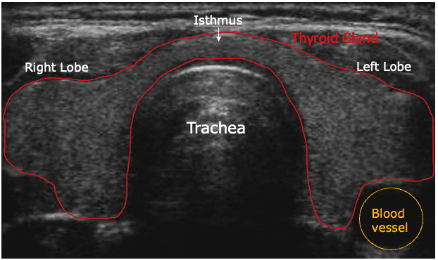
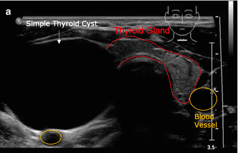
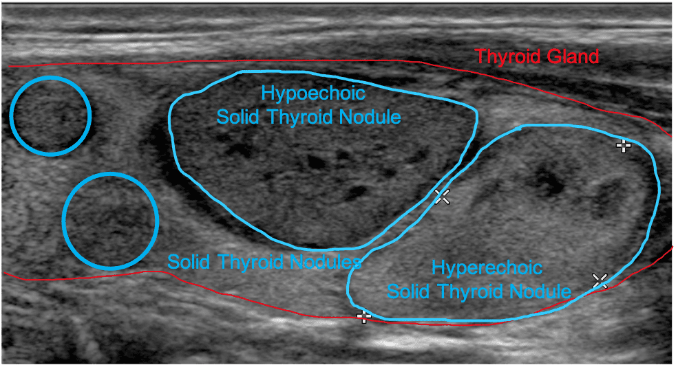
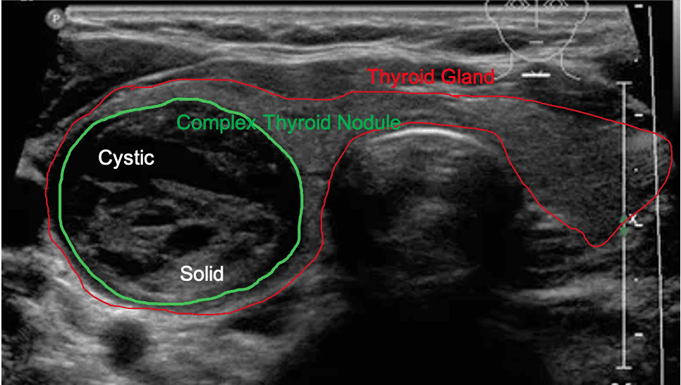
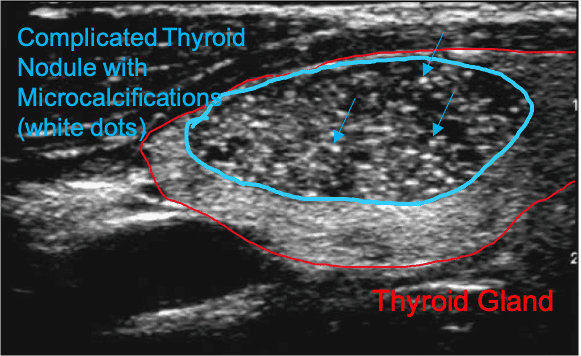
You’ve made it to the final episode of the Thyroid Ultrasound Trilogy! Give yourself a pat on the back. In this article, we will guide you through some basic sonographic features of your thyroid on ultrasound. The thyroid gland belongs to the endocrine family. It is responsible for the production of thyroid hormones to maintain essential bodily functions. The thyroid is situated in the anteroinferior (front and lower) part of the neck, just below your larynx (Adam’s apple). It consists of right and left lobes located along either side of the trachea (windpipe). The two lobes are connected by a narrow bridge of tissue called the isthmus, across the midline, draping over the anterior tracheal wall. The parathyroid glands, usually four in number, lie on the posterior side of the (on the back) thyroid gland, two on each side.

Normal Thyroid on Ultrasound

On ultrasound, the normal thyroid gland has a homogeneous (smooth/uniform), medium- to high-level echogenicity (brightness level). Key landmarks include the trachea, esophagus, blood vessels (carotid arteries and jugular vein), and surrounding muscle groups. The size and shape of the thyroid lobes vary widely in the normal population. In adults, the mean length is 40mm-60mm, and the mean thickness of the lateral lobes is 13mm-18mm. When the thickness is greater than 20mm, the thyroid gland may be considered enlarged.

Thyroid gland in the Transverse plane
Thyroid gland in the Sagittal plane

Thyroid Nodules on Ultrasound

Nodules, which are overgrowths on the thyroid can exhibit a range of characteristics including cystic (fluid-filled), solid (tissue-filled), complex (cyst + solid), and calcifications (calcium deposits).
Cystic nodules generally have a low possibility of being cancerous. Simple thyroid cysts appear anechoic (black) on ultrasound as opposed to complex cysts appear with low-level internal echoes reflected from particles within the cyst.

thyroid cyst
Thyroid gland in the Transverse plane
soild thyroid
Thyroid gland in the Sagittal plane

Solid nodules have a higher chance than cystic nodules of being cancerous. But solid nodules are common entities in thyroid glands and a large majority of them remain benign throughout their course. They can appear hyperechoic (brighter) or hypoechoic (darker) on ultrasound depending on its tissue composition.

Complex nodules ontain both fluid and solid components, hence, “complex” in composition and appearance. Cystic parts are generally anechoic (black), sometimes complex cysts can also exhibit echoes in the fluid-filled structure. Solid components are generally more echogenic than the cystic parts.

complex thyroid
Thyroid gland in the Transverse plane
complicated thyroid
Thyroid gland in the Sagittal plane

Calcifications can sometimes be observed in thyroid nodules as little white dots or areas. They usually represent degenerative processes and may represent signs of malignancies that would require the nodule to undergo further investigations such as a biopsy.

You’ve made it to the end of the Thyroid Ultrasound Trilogy! Now you should have a much better understanding of how your thyroid looks on the most commonly used imaging modality to examine your thyroid.

Remember, your doctor is ALWAYS the ultimate go-to person for any of your medical-related questions.

Schedule an appointment with a specialist today to discuss your thyroid issues

Patients should talk to their doctor to decide if STARmed Thyroid RFA is right for them. Patients and doctors should review all available information on non-surgical and surgical options and associated risks in order to make an informed decision. Individual results may vary, testimonials are not claimed to represent typical results. All testimonials are from real patients, may not reflect the typical patient’s experience, and are not intended to represent or guarantee that anyone will achieve the same or similar results. The information on this site is not intended or implied to be a substitute for professional medical advice, diagnosis, or treatment. All content, including text, graphics, images, and information, contained on or available through this website is for general information purposes only.

Sanders, R. C. (2016). Clinical Sonography: A practical guide. Philadelphia: Wolters Kluwer.

Rumack, C. M., Wilson, S. R., & Charboneau, J. W. (2005). Diagnostic ultrasound. St. Louis: Elsevier Mosby.

Leave a Comment

Your email address will not be published. Required fields are marked *

More From Our Blogs

Diagnostic

Thyroid Ultrasound Trilogy – I. The Basics of Ultrasound

Back to blog Have questions about your thyroid ultrasound? We are here to answer! Inspired by some patients’ requests, we have prepared a series of Thyroid Ultrasound articles to help you better understand those terminologies on

Why Might Thyroid Nodules Need To Be Surgically Removed?

Thyroid nodules can be diagnosed as two primary forms, malignant (cancerous) or benign (non-cancerous). This diagnosis is one of the most significant factors considered before a suitable method of treatment is prescribed. Is it necessary

Important Safety Information / Disclaimer
Patients should talk to their doctor to decide if STARmed Thyroid RFA is right for them. Patients and doctors should review all available information on non-surgical and surgical options and associated risks in order to make an informed decision. Individual results may vary, testimonials are not claimed to represent typical results. All testimonials are from real patients, and may not reflect the typical patient’s experience, and are not intended to represent or guarantee that anyone will achieve the same or similar results.
The information on this site is not intended or implied to be a substitute for professional medical advice, diagnosis or treatment. All content, including text, graphics, images and information, contained on or available through this web site is for general information purposes only.
Registered trademarks of TaeWoong Medical USA or their respective owners.

Indication
The VIVA Combo RF Ablation System and star RF Electrode are intended for use in percutaneous and intraoperative coagulation and ablation of tissue.

Contraindications
There is a risk that error may result due to the radiofrequency current on patients who have pacemakers and other active implants. Do not use the radiofrequency lesion generator and electrode on these patients.
Complications:
The following types of complications may result due to the use of the radiofrequency lesion generator and electrode.
– Tumor recurrence
– Burn due to the over-heating of the the surgical equipment
– Dangerous situation due to the unskilled equipment control
– Cross-infection or complications due to the re-use of the inappropriate electrode
– Ascites/diarrhea
– Bleeding of the coagulated part
– Ventricular fibrillation
– Weakness of liver functions
– Symptoms after RFA treatment includes (abdominal) pain, fever, nausea, headache, right shoulder joint pain and chest discomfort might occur

RFA for Life is committed to protecting your privacy under the relevant laws in the USA, and we’ll only use your personal information to administer your account and to provide the products and services you requested from us.
By clicking submit below, you consent to allow RFA for Life to store and process the personal information submitted above to provide you with the contents requested.

Physician Finder

You are taking a big step by exploring your treatment options. You are not alone. Many other people were searching for the same answers too. Many of these individuals chose RFA.