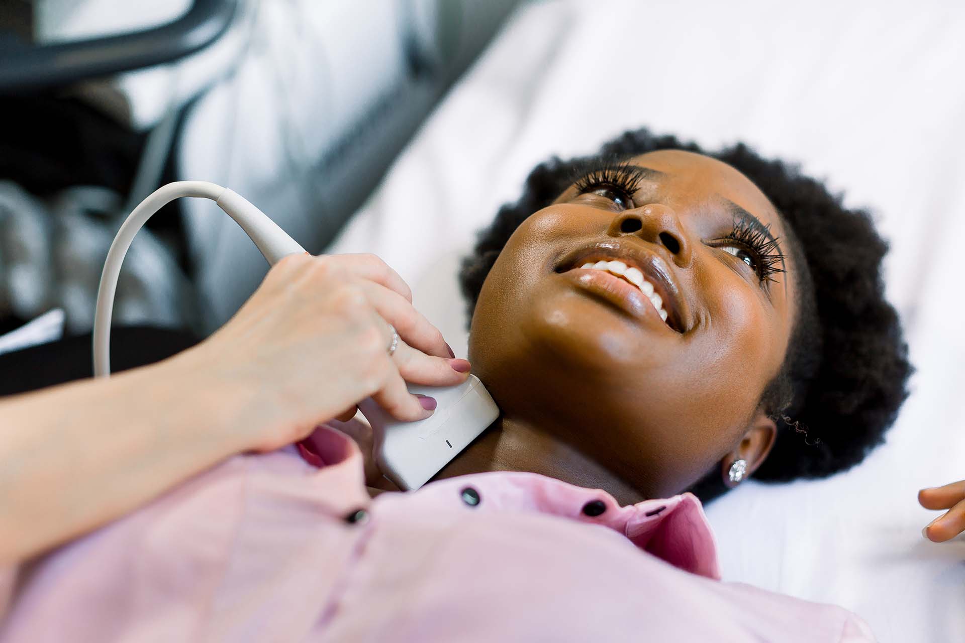
Thyroid nodules are a common medical problem in the USA. While most thyroid nodules (about 85%) are benign and need follow-up, some require treatment for associated symptoms and cosmetic issues.
With technology advancement, many minimally invasive treatments have been introduced to treat thyroid nodules non-surgically under ultrasound guidance:
- Percutaneous Ethanol Injection (PEI): recommended for cystic (fluid-filled) thyroid nodules.
- Hyperthermic treatment: Laser Ablation and Radiofrequency Ablation (RFA) recommended for solid (tissue-filled) benign thyroid nodules.
- Others still under investigation: high-intensity focused ultrasound (HIFU), microwaves, cryotherapy, electroporation
This article focuses on detailing the basic principles, indications, and techniques designed to optimize the thyroid RFA procedure along with its clinical results and expected potential risks.
This summary aims to extract the most relevant information from the clinical study for patient education. If you are interested inreading the full clinical study, click here.
Principle of Radiofrequency ablation
RFA burns (ablates) the targeted nodular tissue with thermal (heat) energy produced from high-frequency electric waves ranging from 200 to 1200 kHz. During the procedure, a radiofrequency generator that creates high-frequency electric waves is connected to an electrode inserted directly into the thyroid tumor, delivering energy to the targeted area.
By disturbing the tissue located within a few millimeters around the electrode, heat energy is produced to ablate the tumor tissue immediately next to the electrode, causing destruction within the area. In addition to the direct heat effect, heat conduction from the ablated area can result in relatively slower damage to the tissue further away from the electrode tip. The extent of heat created is controlled by the specialist carrying out the procedure. The efficacy of this procedure depends on the temperature used and the composition of the tissue being treated.
Indications and Patient Selection
Generally, patient selection follows the criteria laid out in the Koran Society of Thyroid Radiology (KSThR) guideline, which was first established in 2009 then further revised in 2012 and 2017. Thyroid RFA is mostly recommended for patients with benign thyroid nodules who have cosmetic concerns or symptoms.
American Thyroid Association (ATA) guidelines also recognized Radiofrequency ablation(RFA) to be performed for certain recurrent thyroid cancers for patients at high surgical risks or who refuse surgery.
Pre-Procedure Assessment
Various precautions are taken both before and after thyroid Radiofrequency ablation(RFA) to ensure safety and efficacy.
Before thyroid RFA, several assessments are performed:
- Fine needle aspirations to confirm benign thyroid nodules. Fine needle aspiration (FNA) biopsy is a process of simply removing some cells or fluid from the affected area using a fine needle for examination under a microscope.
- Subjective assessment of symptoms of thyroid nodules and cosmetic issues
- Ultrasound examinations to assess characteristics of thyroid nodules and to confirm benign features of cytologically proven thyroid nodules.
- Blood tests and laboratory tests of serum hormone levels to evaluate the success of any ablation.
During The Procedure
Thyroid
RFA can be performed at the radiology unit or in the operating room.
It is performed
under continuous ultrasound (US) monitoring to provide a clear
visualization of the thyroid nodule.
The procedure should be performed by a surgeon or a radiologist experienced with thyroid US, Fine Needle Aspiration (FNA), and RFA. Before the procedure, patients will receive local anesthesia (1-2% lidocaine hydrochloride) at the puncture site. Some surgeons prefer to perform RFA without general anesthesia for the early detection of complications. Following the treatment, patients should be kept under observation for 1-2 hours.
Follow-up
Similarly,
after the procedure, several tests need to be performed to measure
the treatment efficacy. Evaluations include:
- Clinical
and cosmetic symptoms of thyroid nodules - Complications
- Thyroid
nodule volume - Follow-up
clinical examinations at 1, 3, 6, and 12 months and each year for up
to 5 years.
Therapeutic success has been defined as a 50% volume reduction at 12 months. Additional treatment will be recommended if follow-up ultrasound shows a remaining viable portion of the nodule and if the patient complains of ongoing clinical or cosmetic symptoms.
Risks And Adverse Events
According
to a recent meta-analysis with 728 nodules in 715 patients, overall
and major complication rates
were 3.2% and 0.7%, respectively.
| Minor Complication |
Major Complication |
| Pain | Voice Change |
| Hematoma | Nodule Rupture |
| Vomiting | Permanent hypothyroidism |
| Skin burns |
Brachial plexus injury |
| Transient Thyroiditis |
The overall incidence for voice change (transient and permanent) is 1.44%.
Incidence of voice change was higher in a patient with recurrent thyroid cancer (7.95%) than in patients with benign thyroid nodule (0.94%)
Conclusion
The study shows favorable outcomes with RFA in thyroid nodule management regarding size reduction, toxic nodule cure, and compressive symptom resolution. Future studies are needed to guide the use of this novel technique as an alternative treatment option for thyroid diseases and thyroid cancer.


