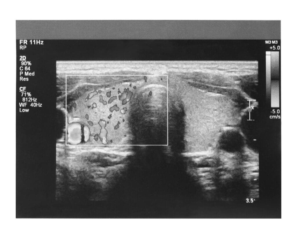
Summary: A thyroid ultrasound is a quick, non-invasive procedure that helps examine your thyroid. Knowing basic ultrasound medical terms can ease anxiety and improve communication with your doctor. The blog explains terms like ultrasound gel, sonogram, transducer, echogenicity, and thyroid-specific terms such as cysts and nodules.
A thyroid ultrasound, also called a thyroid sonogram or thyroid echogram, is a non-invasive exam. It’s a brief, painless procedure that allows your physician to examine your thyroid gland. It’s a common, effective, and fast way for your doctor to see a detailed image of your thyroid. It can be used as a diagnostic tool or to guide an electrode during your RFA procedure.
If you’re preparing for your first thyroid ultrasound, you may be feeling nervous. We’ve found it’s empowering to learn a few basic ultrasound medical terms before your test. It can help you understand the exam and improve communication with your doctor. Having the right language is particularly important if you’re feeling stressed about the exam or results.
Below, you’ll find a simple glossary of ultrasound medical terminology, including some thyroid-specific ultrasound terms. Continue reading to empower yourself before your thyroid exam.
Basic Ultrasound Medical Terms
Ultrasound Gel (or Couplant Gel)
During the test, your physician will place some ultrasound gel on your neck. This is the medium that the soundwaves pass through to create the ultrasound images. It also helps the hand-held transducer glide smoothly over the skin.
Ultrasound gel is primarily made of water. It is fragrance-free and will not stain or damage skin or clothing.
The gel may be somewhat cold, but doctors often warm the gel before an exam of the thyroid. You can easily remove the gel with a tissue or towel after your ultrasound.
Sonogram
A sonogram is the name for the image generated during your ultrasound. Sonography is a medical term interchangeable with “ultrasound.”
During diagnostic exams, the sonographer or the ultrasound technologist will most likely capture sonogram images digitally. At some practices, your doctor may print out a sonogram to include in a physical medical file.
Sound Waves
The ultrasound exam creates sonogram images using sound waves. These are invisible energy waves that pass through the ultrasound gel and your skin to create images. The structures in your body reflect the sound waves to create echoes.
Transducer
The transducer is the hand-held part of the ultrasound machine. This is the part of the machine that generates sound waves. Your doctor will hold the transducer to your neck to create and capture sonogram images during your exam. While you might feel some light pressure on your neck, the transducer is smooth and the exam is typically painless.
Echogenicity
Echogenicity is another word for brightness when discussing sonogram images. Some tissue and structures can reflect ultrasound waves. Others cannot. Thus, your doctor can tell the composition of different cysts and nodules based on the echogenicity of the image.
- An echogenic structure will appear brighter than surrounding structures.
- A hyperechoic structure will appear darker than surrounding structures.
- An echo-free structure will appear black on a sonogram. This is usually a sign of a thyroid cyst.
Ultrasound Interpretation
Sometimes, your ultrasound will be performed by your doctor. This will be the case during the RFA procedure. Most of the times, however, a diagnostic ultrasound may be performed by an ultrasound technician.
Legally, technicians cannot provide ultrasound interpretation. That means they cannot share results with you. Instead, they will send your sonogram images to your doctor. Your doctor will interpret the images and meet with you to share the results of the exam.
While this wait can be nerve-wracking, it’s important to be patient to receive the most accurate reading of your images.

Thyroid-Related Ultrasound Terms
Thyroid Ultrasound
Doctors can perform an ultrasound test on many parts of the body. A thyroid ultrasound is an exam of the thyroid gland in your neck. It can help identify cysts, nodules, goiters, tumors, and issues with the lymph nodes near the neck.
Thyroid Cyst
A thyroid cyst is a fluid-filled lump in the thyroid gland. Cysts can vary in size but carry an extremely low possibility of malignancy. On your sonogram, they will appear as a black (echo-free), round structure in the thyroid.
Thyroid Nodule
While all cysts are nodules, not all thyroid nodules are cysts. “Nodule” is a term for any growth in the thyroid gland. These can include fluid-filled or tissue-filled growths. If a nodule contains both fluid and tissue, it may be called a complex cyst.
Solid, tissue-filled nodules do not appear as dark as cysts on sonogram images. They can vary in appearance. Solid nodules carry a slightly higher possibility of malignancy compared to cysts. Your doctor may follow up with an aspiration test to determine the composition of your nodule.
Discuss These Ultrasound Medical Terms with Your Doctor
Understanding the basic terminology of the ultrasound exam can make the experience less nerve-wracking for patients. Rest assured that this exam is quick, typically painless, and often the first step toward treatment.
Your doctor will most likely use ultrasound technology during the Thyroid RFA procedure. They will use the ultrasound image to locate the structure in your thyroid that needs treatment. Then, they will be able to safely and accurately treat your thyroid under ultrasound guidance. By that point, you will feel comfortable and confident with this safe, non-invasive imaging tool.
Are you wondering if Thyroid RFA might be right for you? Connect with a Thyroid RFA doctor today.


