Previously, we explored the basic physics that powers thyroid ultrasound. In this article, we are going to guide you through some common ultrasound terminologies you might encounter in your thyroid treatment journey.
Brightness (Echogenicity) Terms
- Echogenicity: term used to describe the ability of a structure to reflect ultrasound waves and bounce (generate) echoes. The brightness of a structure appears on ultrasound screen depends on its surround structures.
- Hyperechoic (More Echogenic): structure appears brighter (more echogenic) on ultrasound than surrounding structures.
- Hypoechoic (Less Echogenic/Echopenic): structure appears darker (less echogenic or echopenic) than surrounding structures.
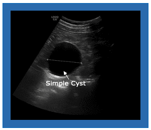
- Anechoic/Echo-free: structure appears black, no internal echoes are produced (bounced) from the structure. Typically see this in simple fluid-filled cysts.
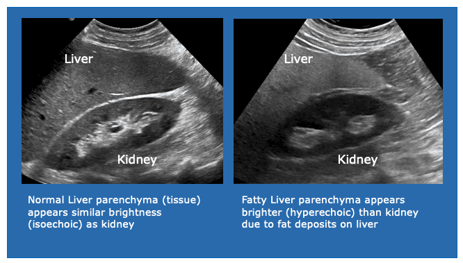
- Isoechoic: structure exhibits the same brightness as its surround structure, same echogenicity.
- Echogenicity terms are relative. Ex. The normal liver parenchyma appears isoechoic to kidney’s echotexture. However, when liver becomes fatty, it appears hyperechoic or more echogenic than the kidney.
Characteristic Terms
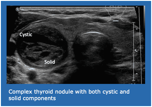
Cyst: Well-defined, spherical, fluid-filled structure with sharp/clear back wall. Contains few or no internal echoes. Simple cyst usually appears anechoic on ultrasound. Cystic is used to describe fluid-filled structures. Cystic nodules usually carry a low possibility of malignancy.
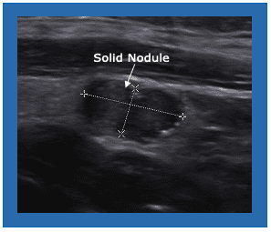
Simple: a structure that has only one composition either solid (echogenic) or fluid-filled (anechoic).
Complex: a structure that contains both solid and fluid-filled components.
Now you’ve learned some common terminologies used in ultrasound. In the next article, we will go through some basic sonographic features of the thyroid.
Remember, your doctor is ALWAYS the ultimate go-to person for any of your medical-related questions.
Patients should talk to their doctor to decide if STARMED Thyroid RFA is right for them. Patients and doctors should review all available information on non-surgical and surgical options and associated risks in order to make an informed decision. Individual results may vary, testimonials are not claimed to represent typical results. All testimonials are from real patients, and may not reflect the typical patient’s experience, and are not intended to represent or guarantee that anyone will achieve the same or similar results. The information on this site is not intended or implied to be a substitute for professional medical advice, diagnosis or treatment. All content, including text, graphics, images, and information, contained on or available through this website is for general information purposes only.


10 Responses
It’s interesting I wish to have more learning session on my email including picture and practice video
I need to learn more
Perfect.
Thank you.
I’m interested in receiving further sonography education, including visual aids and video demonstrations, through email.
Great
I love this
Nice understanding
I was suggested this blog by my cousin. I’m not sure
whether this post iis written by him as no one else know such detailed about my trouble.
You are incredible! Thanks! https://menbehealth.Wordpress.com
This is very educational
You are so awesome! I do not suppose I have read anything like this before. So wonderful to discover somebody with original thoughts on this subject matter. Really.. thanks for starting this up. This web site is one thing that’s needed on the internet, someone with some originality!